
Cells are the fundamental building blocks of life, yet the intricacies of their internal architectures have long been shrouded in mystery due to limitations in imaging technologies. Traditional optical microscopes have struggled to capture the minute details within cells, which are often obscured by the constraints of their resolution capabilities. Observing cellular structures with precision is essential for advancing our understanding of biological processes, and recent breakthroughs in microscopy have opened new avenues for scientific exploration.
In a collaborative effort between researchers at the Universities of Göttingen and Oxford, alongside the University Medical Center Göttingen (UMG), a pioneering microscopic innovation has emerged that possesses extraordinary resolution capabilities of less than five nanometers—an achievement that surpasses previous standards significantly. To put this achievement into perspective, consider the incredible scale: five nanometers can be likened to a hair strand being subdivided into 10,000 segments.
Historically, conventional microscopes could only resolve details beginning around 200 nanometers, which left many crucial cellular structures obscured or poorly defined. For instance, the cytoskeletal frameworks within human cells comprise tubes measuring a mere seven nanometers in diameter. And for the synaptic cleft, the narrow gap allowing communication between two neurons, the dimensions hover between 10 to 50 nanometers. The limitations of prior technologies rendered the visualization of these essential features virtually impossible. However, the newly developed microscope promises to bridge this gap, yielding images that are richly informative and rich in detail.
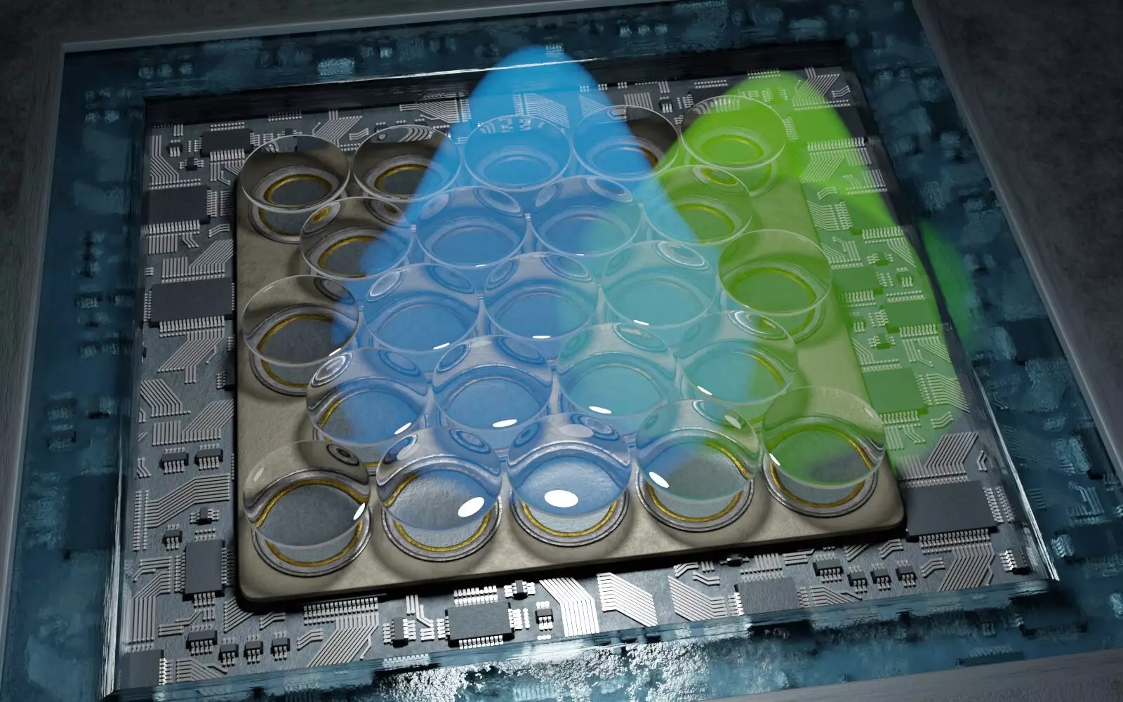
The innovative microscope functions on principles of fluorescence microscopy and employs “single-molecule localization microscopy” (SMLM). This technique involves the precise switching on and off of individual fluorescent molecules within a sample, allowing researchers to map their exact positions. Collectively, these localized points can be reconstructed into a comprehensive model of the structure being studied. Prior to this advancement, resolutions hovered between 10 to 20 nanometers. The research group led by Professor Jörg Enderlein at Göttingen has effectively doubled this resolution, exemplifying a significant leap in the technology’s capabilities.
Enderlein describes this advancement as a groundbreaking milestone in high-resolution microscopy. One of the key advantages of the new method is its cost-effectiveness and ease of use compared to alternative imaging technologies. Such accessibility is crucial for fostering wider adoption within the scientific community, enabling more researchers to probe the complex architectures of cells.
The implications of this innovative microscopy technique are manifold. By facilitating the visualization of minuscule cellular components, researchers can glean insights into cellular organization, functionality, and the molecular interactions that underpin life processes. This could potentially transform fields like neurobiology, where understanding synaptic connections and protein organizations can shed light on brain function and the mechanisms underlying neurodegenerative diseases.
Moreover, the team has developed an open-source software suite to streamline the analysis of the data generated by this novel microscopy. This democratization of technology ensures that scientists across various disciplines can benefit from enhanced imaging capabilities, leading to collaborative investigations and a deeper understanding of biological phenomena.
The advent of this advanced microscopy technique marks a significant turning point in our ability to visualize the intricate details of cellular structures. As we delve deeper into the microscopic world, the potential for discoveries that can reshape our understanding of biology is vast. The clarity finally offered by these new tools not only pushes the boundaries of scientific research but also democratizes access to advanced imaging technology, setting the stage for a new era of exploration in the life sciences. As we expand our understanding of cellular mechanics and structures, the pathways to innovations in medicine, biotechnology, and fundamental biology become clearer than ever.