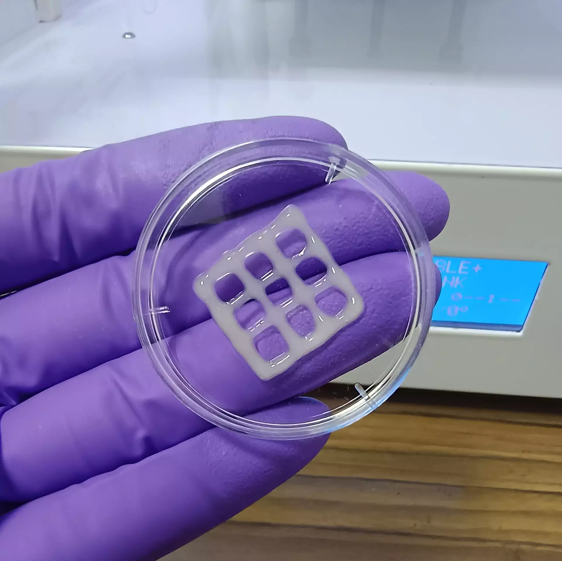
Lung diseases pose a significant threat to global health, contributing to millions of deaths annually. Despite advancements in medical science, the treatment landscape remains constrained, with limited options available for patients suffering from chronic conditions like chronic obstructive pulmonary disease (COPD) and cystic fibrosis. The quest for new therapeutic strategies continues, but traditional animal models often fall short in mimicking human lung pathology effectively. Recently, researchers have made strides in biotechnology, creating a mucus-based bioink designed for 3D printing of lung tissue. This new development promises to unlock possibilities for better understanding and managing lung diseases.
The statistics surrounding lung diseases are alarming. Millions succumb to conditions that compromise respiratory function, and while some individuals benefit from lung transplants, the scarcity of donor organs is a significant barrier. Medications can alleviate symptoms but often fail to provide a definitive cure. For diseases like cystic fibrosis and COPD, where long-term management is essential, finding effective treatments has proven arduous. Traditionally, researchers have relied on rodent models to test potential pharmaceuticals; however, these models are known to inadequately represent the complexities of human lung diseases.
In recent years, bioengineering has emerged as a groundbreaking field with the potential to address these challenges through the production of artificial lung tissue. One promising approach involves 3D printing, which enables the creation of tissue models that closely mirror human anatomy. Nonetheless, the successful implementation of this technique hinges on the availability of suitable bioinks that can support cellular proliferation and mimic the biochemical characteristics of actual lung tissue.
The research team led by Ashok Raichur recognized the potential of mucin, a component of mucus that has yet to be fully utilized in bioprinting. Mucin contains structural elements reminiscent of epidermal growth factor, an essential protein involved in cell growth and adhesion. By chemically modifying mucin into a methacrylated form known as MuMA, the researchers aimed to create an environment conducive to lung cell growth.
The process to develop the bioink involved several innovative steps. To enhance the viscosity of MuMA and promote better cell adhesion, researchers incorporated hyaluronic acid—an existing natural polymer renowned for supporting tissue structure. The resulting bioink was tested through a series of 3D printed structures characterized by various geometric patterns, such as grids.
Upon exposure to blue light, a crosslinking reaction occurred which formed a stable porous gel. This crosslinked structure, touted for its ability to retain moisture, created an excellent microenvironment for cell survival and proliferation. The interconnected pores within the gel facilitate the diffusion of oxygen and nutrients, essential for encouraging the development of respiratory tissue.
Critically, the printed bioink structures demonstrated nontoxic properties and a gradual biodegradation rate, positioning them as potentially viable implants for future therapeutic applications. As these bioinks degrade, they could be supplanted by new lung tissue, cultivated from the patient’s own cells, thus minimizing issues of rejection and structural compatibility.
Additionally, the advancements introduced by this research extend beyond mere tissue replacement. The bioink could serve as a groundbreaking platform for constructing 3D lung models to study disease mechanisms and explore innovative treatment avenues. As investigations proceed, these models could usher in a new era of personalized medicine where therapies are tailor-fitted to individual pathologies of lung disease.
The successful creation of mucus-based bioink for 3D bioprinting offers vital tools in the ongoing battle against lung disease. As researchers continue to refine these techniques, the potential for developing novel treatment modalities grows. This pioneering work represents a hopeful step towards improving the quality of life for patients battling chronic lung diseases and inspires future innovations in tissue engineering and regenerative medicine.

