
A recent study conducted by researchers at the University of California, Los Angeles, has unveiled a groundbreaking advancement in 3D Quantitative Phase Imaging (QPI) through a novel wavelength-multiplexed diffractive optical processor. Traditional QPI techniques, albeit effective, have been notoriously hindered by their reliance on multiple illumination angles and laborious digital post-processing for image reconstruction. The new methodology not only simplifies this process but offers a transformative leap in the way we capture images of transparent specimens. This innovative approach could significantly alter the landscape of biomedical imaging and diagnostic technologies.
Efficiency Meets Precision
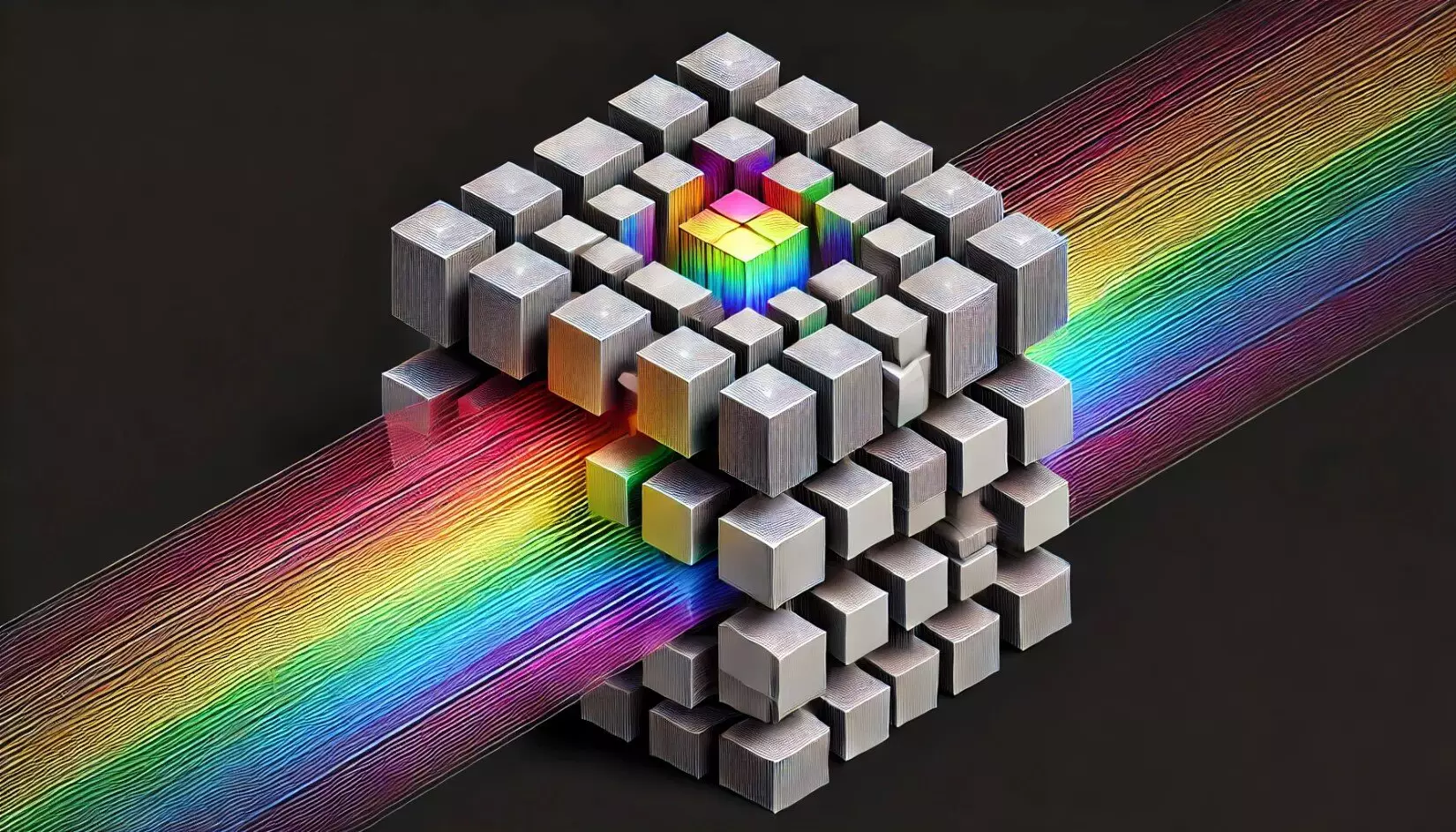
The crux of the innovation lies in the processor’s ability to translate phase distributions of various two-dimensional objects located at different axial planes into intensity patterns, each encoded within its unique wavelength channel. This sophisticated mechanism permits the acquisition of detailed phase images utilizing only intensity-based image sensors, effectively bypassing the complications and computational overhead of traditional digital phase recovery techniques. Aydogan Ozcan, the lead researcher and a prominent figure in the field, expressed optimism regarding the implications this technology holds for biomedical applications, highlighting the promise of high-resolution, label-free imaging that could advance clinical diagnostics and research.
Deep Learning Optimization
This state-of-the-art system capitalizes on the synergy between wavelength multiplexing and passive diffractive optical components, meticulously optimized through deep learning algorithms. This is where the true genius of the design shines; it is not merely about imaging, but also about how efficiently phase-to-intensity transformations can occur even across multiple axial planes. The rapid pace at which quantitative phase imaging can be performed opens up new avenues for research, ushering in a future where real-time imaging and diagnostics could become a norm rather than an exception.
Expanded Utility Across Spectrums
Validation through proof-of-concept experiments demonstrated the processor’s efficacy in imaging distinguishable phase objects at varying axial positions within the terahertz spectrum. Moreover, this technology showcases versatility owing to its adaptable nature, with potential expansions into other segments of the electromagnetic spectrum, such as the visible and infrared ranges. This adaptability paves the way for pioneering developments in integrated imaging systems that can be portable and efficient—perfect for applications in environmental monitoring, materials science, and more.
A Paradigm Shift for Biomedical Applications
The implications of this research extend far beyond mere imaging. The streamlined process of achieving high-quality QPI images offers unparalleled opportunities for improved disease diagnostics, materials characterization, and even environmental sample monitoring. In a world where the speed of diagnosis and research is crucial, this new methodology could accelerate our capabilities in understanding diseases and materials at their very core. By harnessing advanced technologies and innovative designs, we may very well be stepping into a new era of precision and efficiency that will redefine our abilities in scientific exploration and healthcare advancements.