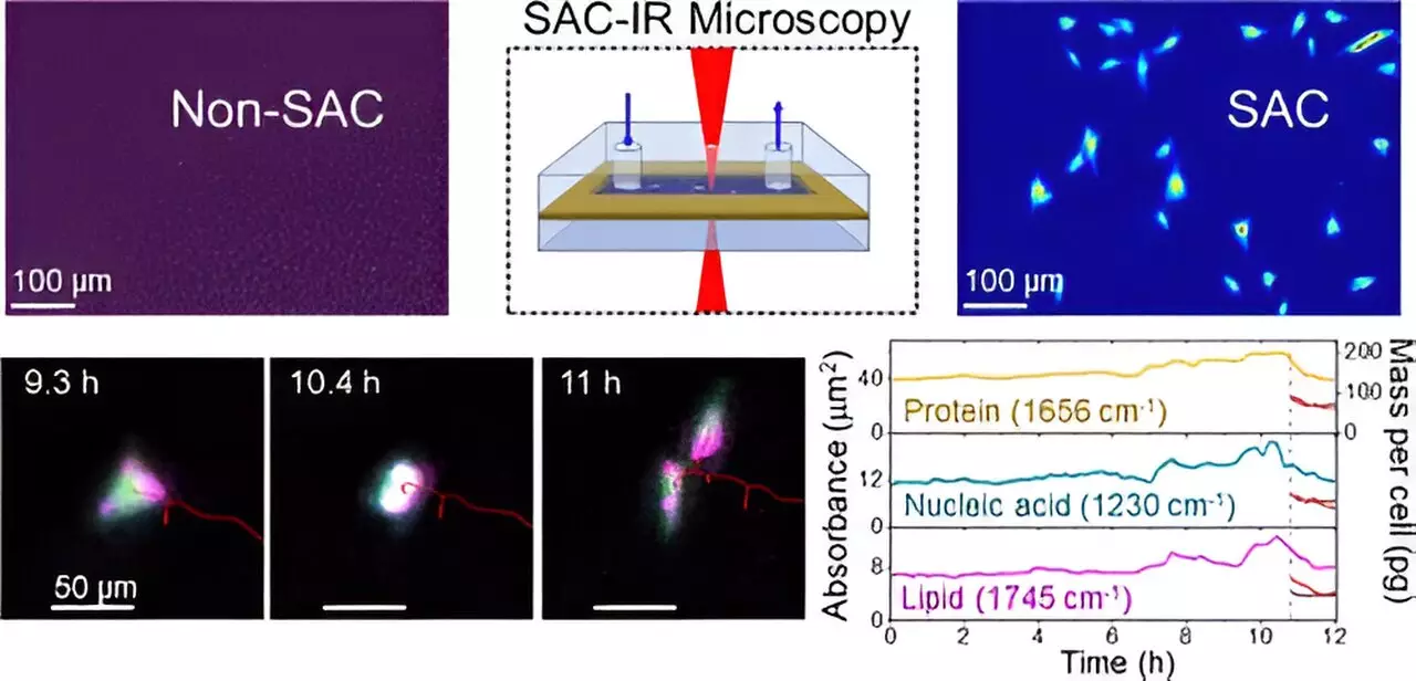
As the global landscape of biotechnology continues to expand, the demand for innovative techniques to analyze biomolecules within living cells is more pressing than ever. These developments are crucial for multiple applications, including drug therapies and biomanufacturing. Recently, groundbreaking research from the National Institute of Standards and Technology (NIST) has introduced a novel method leveraging infrared (IR) light for this purpose. Unlike traditional methods limited by the absorptive properties of water in cells, this advancement heralds a new era in quantitative biomolecule imaging, which could accelerate scientific progress in various fields.
The Challenge of Water Absorption in IR Imaging
Understanding the role of water in biochemistry is essential. While water is ubiquitous in cellular environments, its unique properties pose a significant obstacle in IR microscopy. Water has a strong tendency to absorb IR radiation, which obscures the absorption signals from other biomolecules. This can generate an “optical masking” effect, making it nearly impossible to accurately observe the proteins, lipids, and nucleic acids essential for fundamental biological processes.
To illustrate this problem, one can liken it to spotting an airplane against a bright sunlit sky—without the right tools, the airplane goes unnoticed. Researchers, particularly NIST chemist Young Jong Lee, recognized that an innovative solution was required to “unmask” the signals from biomolecules by compensating for water absorption.
Lee’s solution to this challenge manifests as the patented technique called Solvent Absorption Compensation (SAC). This breakthrough method allows scientists to effectively filter out the interference of water, revealing otherwise hidden signals from vital biomolecules. By integrating SAC with a custom-built IR laser microscope, NIST researchers successfully imaged fibroblast cells—cells that are integral to the formation and sustenance of connective tissue. Over a continuous 12-hour observation period, researchers tracked the dynamics of biomolecule clusters during various phases of the cell cycle, including cell division.
This method represents a significant leap forward in comparison to existing technologies that often rely on large synchrotron facilities. In doing so, SAC also preserves the integrity of live cells by eliminating the need for hazardous fluorescent dyes or markers, thereby ensuring consistent and reliable results across laboratories.
The ramifications of SAC-IR extend far beyond mere observation. The method not only quantifies the absolute mass of biomolecules, such as proteins, nucleic acids, and carbohydrates, but also establishes a framework for standardized measurement techniques. In practical terms, this capability has direct implications for a variety of sectors in biomedicine and biotechnology.
One particularly notable application lies in cancer cell therapy. Modified immune cells are often reintroduced into patients with the hope of enhancing their ability to identify and combat cancerous cells. Understanding the safety and efficacy of such modified cells is paramount; thus, SAC-IR can provide critical insights into the biomolecular changes occurring within these cells before they are administered back into the patient.
Future Directions and Research Prospects
Emboldened by initial findings, researchers are eager to explore further applications of the SAC-IR technique. Future studies aim to enhance its accuracy, particularly in assessing other crucial biomolecules, including RNA and DNA. Furthermore, the implications of this research could offer profound insights into fundamental questions surrounding cellular viability. For instance, it may help establish biomolecular signatures that discern living, dying, or dead cells, which is vital for optimizing cell preservation methods.
Another promising avenue includes drug screening and development. By quantitatively analyzing the responses of various cell types to new drug candidates, researchers could identify potential therapeutic agents more efficiently. This could prove transformative in healthcare by expediting the process of drug discovery and assessment, ultimately leading to quicker treatments for patients.
The recent advancement in IR imaging through NIST’s SAC-IR method marks a pivotal moment in biotechnology and cell biology. By overcoming a significant barrier to biomolecular observation, this innovative approach not only enhances our understanding of cellular functions but holds the potential to revolutionize applications in medicine and therapeutics. As researchers refine and expand upon this technique, it promises to illuminate the intricacies of life at the molecular level, paving the way for groundbreaking discoveries in the years to come.

