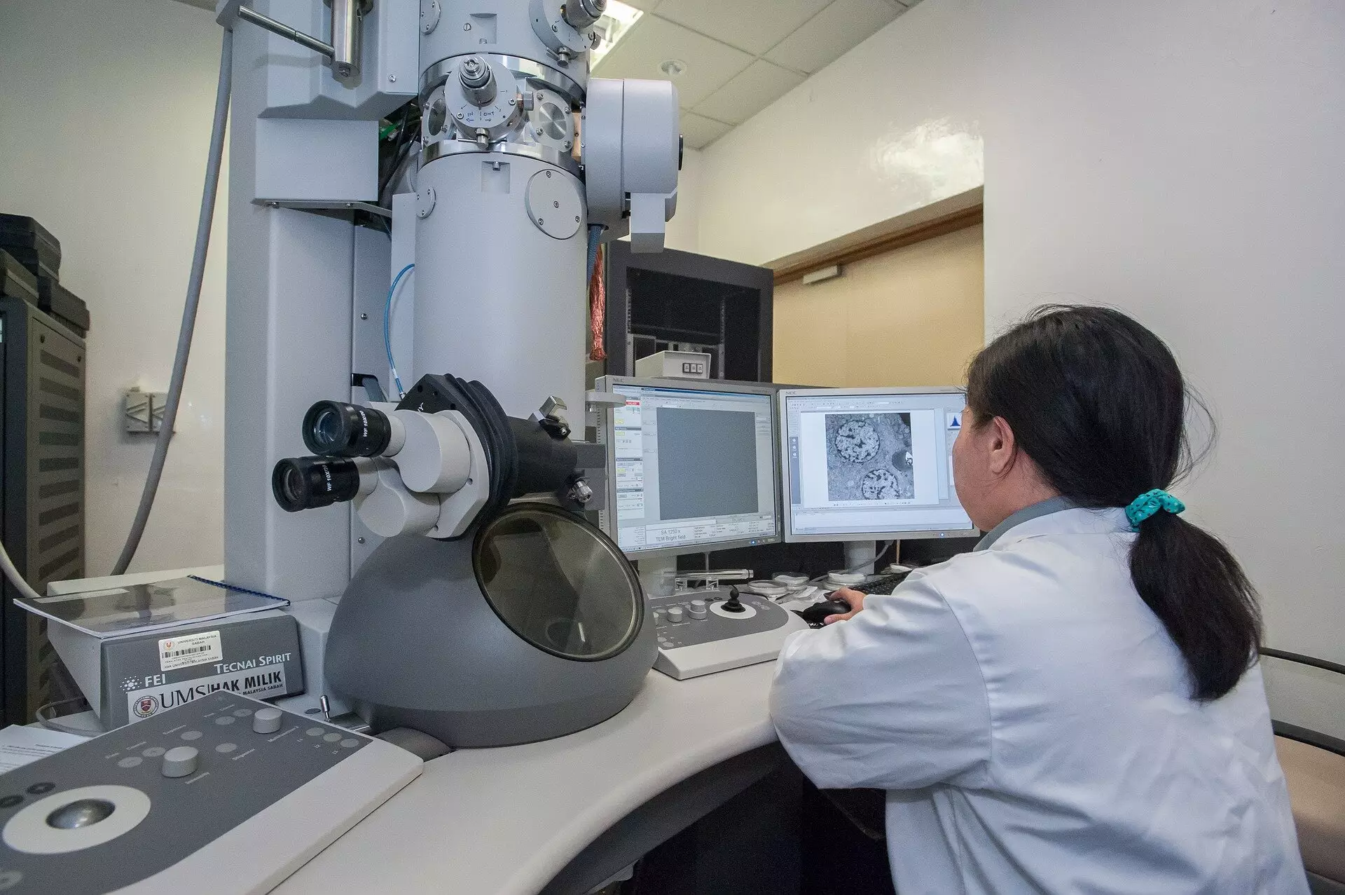
In a monumental advancement that intertwines the domains of materials science and medical imaging, a team of researchers from Trinity College Dublin has developed a pioneering imaging approach that can dramatically cut down both the time and radiation exposure typically associated with electron microscopy. This innovative methodology not only enhances the quality of the images produced but also preserves the integrity of sensitive biological tissues, a crucial factor in the ongoing challenges posed in the field of microscopy. As various scientific domains continue to evolve, the implications of this cutting-edge technology could reshape how we examine and understand materials at the microscopic level.
Understanding Conventional Imaging Limitations
Historically, scanning transmission electron microscopes (STEMs) have employed a methodical scanning of samples through a focused electron beam. This time-tested approach features a predetermined duration during which electrons are directed toward each point, accumulating signals much like traditional cameras taking a still photograph. However, this uniform exposure time, while straightforward, leads to excessive radiation exposure, risking significant damage to the delicate samples under review. The conventional model relies on a principle whereby each pixel gathers electrons until a defined dwell-time elapses. This blind adherence to fixed timing inadvertently subjects sensitive materials to greater risk, potentially compromising the quality of the images obtained and leading to misleading results. The novel imaging technique developed by the Trinity team challenges these long-standing conventions, proposing a more efficient alternative.
The Event-Based Detection System: A Shift in Paradigm
At the core of this revolutionary imaging method lies an event-based detection system that redefines how images are constructed. Rather than adhering to a rigid time frame per pixel, this system captures events as they occur, allowing researchers to measure the time taken to detect a predetermined number of electrons at each sampling point. Through the implementation of this approach, the Trinity College team uncovered a critical insight: the very first electron detected carries substantial information value in constructing the image, while subsequent detections yield diminishing returns. By leveraging this understanding, the team has unlocked a method that not only reduces the number of electrons required for high-quality imaging but also minimizes the damaging radiation exposure traditionally associated with microscopy.
Practical Application and Technological Innovation
Realizing the theoretical foundation into practical usage necessitated the development of patented technology known as Tempo STEM, in collaboration with IDES Ltd. This innovation integrates a sophisticated beam blanker that can effectively “shutter” the electron beam once the requisite image precision has been met. In practical terms, this means that microscopists can now operate with a level of precision unattainable in prior methodologies. As articulated by Dr. Lewys Jones, a leading figure in this breakthrough, the ability to manipulate the illumination in real time opens a new frontier in electron microscopy applications.
Implications for Scientific Research and Medical Imaging
The ramifications of this advancement echo across numerous fields where imaging accuracy is paramount, particularly within biology and sensitive materials research. For instance, when working with biological samples— which are frequently at risk of damage from traditional electron beams—this method could herald a new era of clearer and more reliable data without the catastrophic drawbacks of previous techniques. Dr. Jon Peters emphasizes that while electrons are generally perceived as low-risk concerning radiation, their high-speed impact on delicate samples can lead to significant structural alterations or render them unmeasurable. Therefore, adopting this new imaging strategy is vital for pushing the boundaries of what is possible in research environments.
A Vision for Future Developments
As the scientific community continues to explore the boundaries of discovery, the timing of this advancement could not be more crucial. The reduced radiation exposure alongside improved image quality positions this technology not just as a tool for refinement but as a potential game changer for multiple disciplines. With the ever-increasing complexity of modern materials and biological systems, this innovative approach could facilitate unprecedented explorations into the microscopic unknown.
The development of this advanced imaging technique marks a critical milestone for scientific inquiry, enabling researchers to unravel complexities with increased clarity and confidence. The convergence of innovative technology and profound insight presents exciting opportunities for the future of electron microscopy and, by extension, our understanding of the intricate structures that underpin the natural world.

