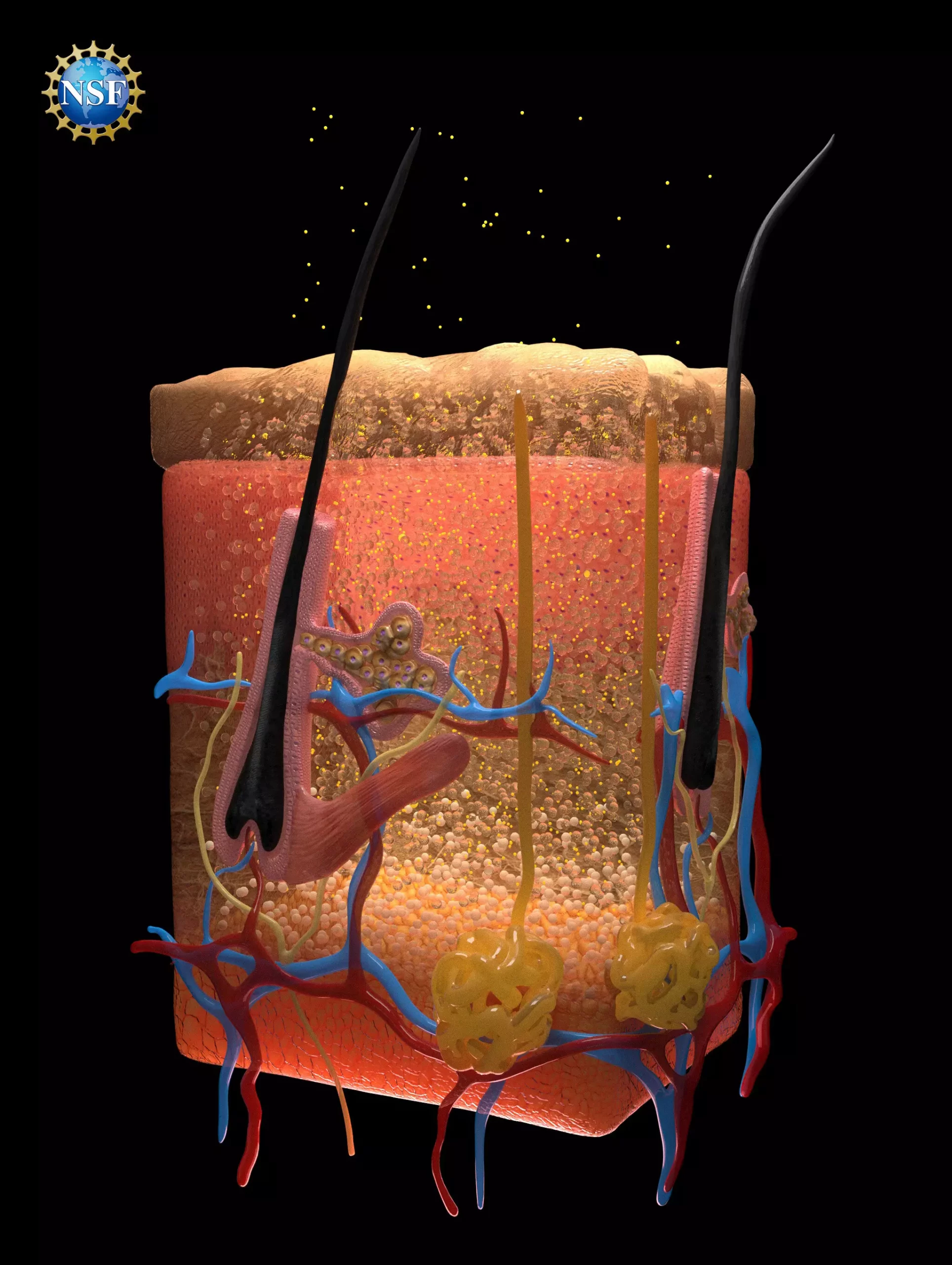
The ability to visualize the internal structures of living organisms has always been a formidable challenge in medical science and biological research. Recent advancements led by researchers at Stanford University are promising to change the landscape of medical diagnostics by enabling a technique to render biological tissues transparent to visible light. This innovative approach could drastically improve the ability to diagnose conditions ranging from traumatic injuries to cancers, paving the way for enhanced treatment plans and patient outcomes.
At the core of this groundbreaking technique lies a seemingly simple yet profound method: the topical application of a food-safe dye. Specifically, the dye used in these experiments is known as tartrazine, or FD & C Yellow 5, commonly found in various foods. The challenge that researchers faced involved understanding how light interacts with differently structured biological materials. For light to travel through biological tissues without scattering, the refractive indices of the dying material and surrounding tissues must be normalized.
Light scattering occurs due to variations in the refractive indices of fats, fluids, proteins, and other components found in tissues. When these components are grouped closely together, they absorb and deflect light in various directions, which leads to the opaque appearance of biological materials. The Stanford researchers recognized that if they could manipulate the refractive indices of these materials through the application of a dye, they could create conditions for light to pass undisturbed, resulting in transparency.
Developing the Technique: A Rigorous Experimental Process
To test their hypothesis, researchers began with tissues from chicken breast. By gradually increasing the concentration of tartrazine, they observed a transition from opacity to transparency as the refractive index of the dye solution matched that of the muscle proteins in the sample. Encouraged by these initial results, the researchers expanded their experimentation to live animal subjects. Upon application of the dye, they achieved remarkable results, rendering skin transparent to reveal underlying blood vessels and internal organ contractions.
The implications of this methodology extend far beyond basic visualization. For example, this transparency could enhance the precision of techniques like laser therapy, particularly in the treatment of cancers and other surface-level conditions. Previously, light penetration was a limiting factor for effective treatment, but the use of this dye could allow lasers to target deeper tissues more effectively.
Impacts on Medical Diagnostics and Future Research
The potential applications for this technology in the field of medical diagnostics are vast. Medical professionals may soon be able to locate vascular injuries more efficiently, evaluate organ health, and even track metabolic processes in real-time, all with a minimal invasiveness. Furthermore, the reversibility of this technique, demonstrated in animal subjects, underscores its viability for practical medical use. The dye’s temporary nature means that once the diagnostic purpose is served, normal tissue opacity can be restored without long-term effects.
Moreover, the foundational research into this technique was not randomly conceived; it evolved through a historical understanding of the interaction between light and matter. By employing principles originally outlined in optics theories from the 1970s and 1980s, which focus on how light can scatter, refract, and absorb, the team was able to make significant strides in biological transparency.
It is essential to acknowledge that the achievement of such innovative scientific breakthroughs often hinges upon the collaborative efforts within research communities. The research team at Stanford began with a small group and eventually expanded to involve over 21 students, advisors, and collaborators. In particular, the inclusion of specialized tools, such as the ellipsometer, has been pivotal in characterizing the optical properties of the dyes used.
This particular instrument, traditionally employed in semiconductor manufacturing, exemplifies how unconventional tools can find relevance in new fields. The shared access to such equipment in institutions fosters an environment ripe for innovation, enabling researchers to arrange resources effectively to achieve groundbreaking discoveries.
The research led by Stanford University researchers represents a transformative advancement in the field of medical imaging. By employing a novel technique to achieve transparency in biological tissues, we may be entering a new era in diagnostics and treatment. This breakthrough could not only enhance our understanding of biological processes but may also lead to earlier detection and more effective treatments for a host of conditions. As continued exploration in this field unfolds, the implications for medicine and healthcare could be profoundly beneficial for society as a whole.

