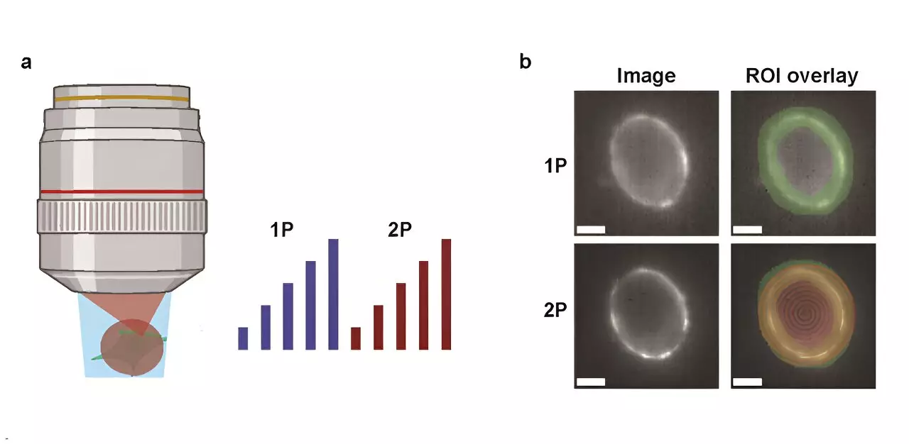
In the evolving landscape of neuroscience, understanding neural communication hinges on the ability to visualize electrical activity in the brain. Recently, genetically encoded voltage indicators (GEVIs) have emerged as pivotal tools in this endeavor. However, the debate surrounding the efficacy of one-photon (1P) versus two-photon (2P) voltage imaging techniques continues to generate interest and scrutiny within the scientific community.
A recent study led by researchers at Harvard University has taken a significant step in clarifying the distinctions between 1P and 2P voltage imaging. Published in the journal Neurophotonics, their research meticulously compares the optical and biophysical limitations inherent to each technique. By evaluating the brightness and voltage sensitivity of commonly utilized GEVIs under both imaging modalities, the research team provides a rich data set essential for future innovation in the field.
The primary goal of this research was to assess how fluorescence diminishes with depth within the mouse brain—a crucial factor for in vivo measurements. The researchers formulated a model that predicts the potential for measurable cell counts based on the properties of voltage reporters, imaging conditions, and the necessary signal-to-noise ratio (SNR). Such modeling represents a valuable contribution, as it aids in strategizing effective imaging approaches.
One of the study’s pivotal outcomes is the revelation that achieving comparable photon count rates in 2P voltage imaging necessitates approximately 10,000 times more illumination power per cell than its 1P counterpart. This staggering requirement raises concerns about tissue photodamage and the accompanying shot noise that may obscure vital data. For instance, when using the JEDI-2P indicator in a mouse cortex, under stringent imaging parameters, the capability to record from only about 12 neurons at depths beyond 300 micrometers highlights the limitations posed by 2P imaging methods.
The implications of these findings are profound. They underscore the inherent challenges in employing 2P voltage imaging for in vivo studies, particularly regarding the need for high SNR while maintaining minimal invasiveness. As current GEVIs present modest voltage sensitivity and stringent photon-count demands, researchers face an uphill battle to achieve comprehensive and deep intercellular recordings.
Looking forward, the necessity for advancements in sensor technology and novel imaging modalities becomes increasingly apparent. The study elucidates that to overcome the limitations of 2P voltage imaging, either substantial improvements to existing GEVIs or entirely new imaging techniques must be developed.
While 1P and 2P voltage imaging techniques each possess unique advantages, the challenges highlighted by this Harvard study reflect the complexities faced in the pursuit of comprehensive neural understanding. Progress in the realm of imaging technologies is crucial to advance our knowledge of neural circuits. As scientists continue to innovate, the insights gleaned from this research will serve as a beacon, illuminating the path toward more effective methodologies in brain imaging.

